
Augmented reality visualization in brain lesions: a prospective randomized controlled evaluation of its potential and current limitations in navigated microneurosurgery
Acta Neurochirurgica (2022) 26:3–14
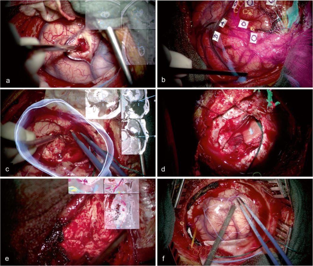
Augmented reality (AR) has the potential to support complex neurosurgical interventions by including visual information seamlessly. This study examines intraoperative visualization parameters and clinical impact of AR in brain tumor surgery.
Methods Fifty-five intracranial lesions, operated either with AR-navigated microscope (n = 39) or conventional neuronavigation (n = 16) after randomization, have been included prospectively. Surgical resection time, duration/type/mode of AR, displayed objects (n, type), pointer-based navigation checks (n), usability of control, quality indicators, and overall surgical usefulness of AR have been assessed.
Results AR display has been used in 44.4% of resection time. Predominant AR type was navigation view (75.7%), followed by target volumes (20.1%). Predominant AR mode was picture-in-picture (PiP) (72.5%), followed by 23.3% overlay display. In 43.6% of cases, vision of important anatomical structures has been partially or entirely blocked by AR information. A total of 7.7% of cases used MRI navigation only, 30.8% used one, 23.1% used two, and 38.5% used three or more object segmentations in AR navigation. A total of 66.7% of surgeons found AR visualization helpful in the individual surgical case. AR depth information and accuracy have been rated acceptable (median 3.0 vs. median 5.0 in conventional neuronavigation). The mean utilization of the navigation pointer was 2.6 /resection hour (AR) vs. 9.7 /resection hour (neuronavigation); navigation effort was significantly reduced in AR (P < 0.001).
Conclusions The main benefit of HUD-based AR visualization in brain tumor surgery is the integrated continuous display allowing for pointer-less navigation. Navigation view (PiP) provides the highest usability while blocking the operative field less frequently. Visualization quality will benefit from improvements in registration accuracy and depth impression.
German clinical trials registration number. DRKS00016955.
The post Augmented reality visualization in brain lesions: a prospective randomized controlled evaluation of its potential and current limitations in navigated microneurosurgery appeared first on Neurosurgery Blog.
This content was originally published here.

